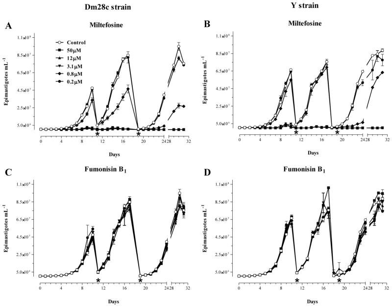
Fig. 2.
Effects of Miltefosine and Fumonisin B1 on the proliferation of epimastigote forms of T. cruzi in vitro. Cultures of epimastigotes (5 × 10⁻⁵.mL−1) of Dm28c (A and C) and Y (B and D) strains were prepared at day 0 in the absence (Control) or presence of increasing amounts of Miltefosine (A, B) and Fumonisin B1 (C, D), and the number of cells was determined daily by direct counting. At days 10 (*) and 19 (*), parasites were collected by centrifugation and adjusted to their starting densities (5 × 10⁻⁵·mL−1) using fresh media containing the same original concentrations of Miltefosine or Fumonisin B1. Each subculture was followed daily by counting for a total of 32 days as indicated at the bottom of each graph. The results shown are the means ± standard error of two sets of independent experiments.

