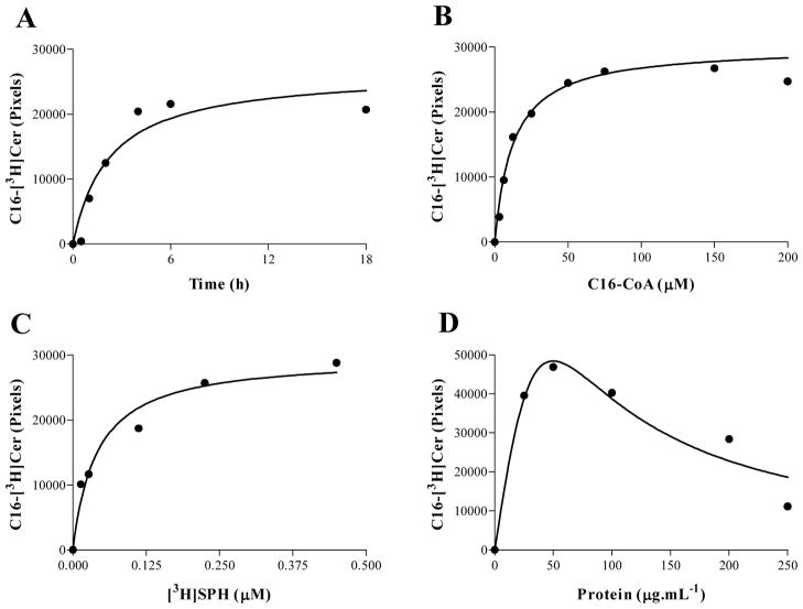
Fig. 5.
Characterization of the T. cruzi microsomal CerS assay. (A) Assays were performed in 100 μL of 100 mM Tris-HCl pH 7.5 containing 1 mM MgCl2, 1 mM DTT, 4 mM NaF, 0.2% (w/v) CHAPS, 0.5 μCi [3H]SPH and 75 μM pamitoyl-CoA at 28 °C for varying periods of time. (B) Measurements were performed as in (A), but all incubations were performed for 4 h; the pamitoyl-CoA concentrations were varied as indicated. (C) Assays were performed as in (B) but with 75 μM pamitoyl-CoA and varied amounts of [3H]SPH. (D) The experiments were performed as in (A) but with incubation for 4 h and varied amounts of microsomes. Lipid extractions and quantification of [3H]Cer were performed as described in item 2.6.

