Fig. 6.
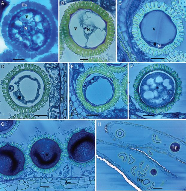
Microgametogenesis in M. esculenta. (A) Early uninucleate microspore. Note dozens of vacuoles (V) with central nucleus (N) forming a star-shape microspore enclosed in mass-synthesized exine (Ex). (B) Mid-uninucleate gametophyte with three vacuoles. Note the centrally located nucleus. (C) Late uninucleate gametophyte with single vacuole. Note the cytoplasm containing nucleus is restricted mostly to the cell periphery. (D) Binucleate gametophyte. A small generative nucleus is located at the periphery whereas the large vegetative nucleus is moving towards the centre along with the cytoplasm. (E) Binucleate gametophyte. Note the vegetative nucleus with two prominent nucleoli. (F) Early deposition of protein and starch in the cytoplasm. Note the generative cell is enclosed in the large vegetative cell. The large vacuole is replaced by the cytoplasm, forming small vacuoles. (G) Male gametophytes completely filled by dense cytoplasm. (H) Mature pollen sac. Note the numerous deformed dead pollen (Dp) grains and live ones (Lp) with dense cytoplasm in the anthers (scale bars A–B = 13.7 µm; C–G = 30.7 µm; H = 126 µm).

