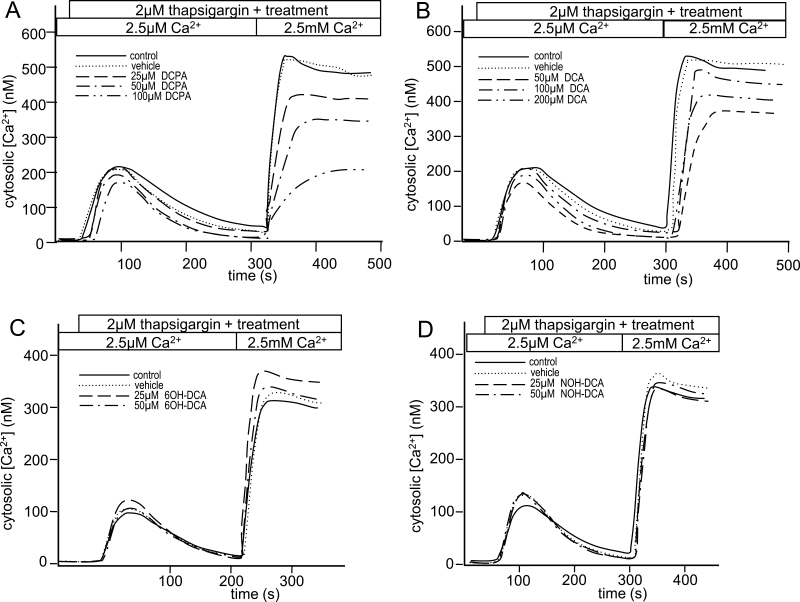
Fig. 4.
Differential effects of DCPA and its metabolites on calcium influx. Jurkat T cells were loaded with the calcium-sensitive dye, fluo-3, and treated with (A) DCPA, (B) DCA, (C) 6OH-DCA, (D) NOH-DCA at concentrations indicated on the figure. Additional treatment groups included a vehicle (veh) control and a no treatment control (labeled “control”). Following addition of the indicated treatment, 2µM thapsigargin was immediately added to deplete ER Ca2+ stores. When fluorescence returned to baseline, 2mM CaCl2 was added and the effect on Ca2+ influx was recorded. Graphs are representative of at least three experiments, and statistical analysis was done collectively on all experimental units (n = 3 for each of DCPA, DCA, and NOH-DCA).

