
Figure 1. PL inhibits proliferation of BL cells in a time- and concentration-dependent manner.
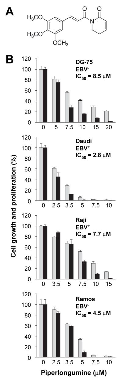
(A) Chemical structure of piperlongumine (PL).
(B) PL-dependent growth inhibition of BL. Tumor cells were seeded in 96-well plates at a density of 1 × 10⁶/ml and grown in presence of the indicated concentration of PL, either for 24 hrs (grey bars) or 48 hrs (black bars). Cell proliferation was measured using the MTS assay. Error bars represent standard deviations of the mean determined in a representative experiment performed in triplicate. IC50 is indicated for the 24-hr treatment. These conditions were used for all experiments presented in Figures 2–5 unless otherwise stated.