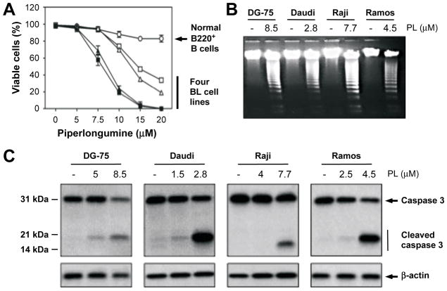
Figure 2. PL-induced apoptosis involves activation of caspase-3.
(A) PL kills BL but not normal B cells. Normal peripheral blood B-lymphocytes obtained by MACS separation using B220 columns (circles) and BL cells (open squares, DG-75; closed squares, Daudi; open triangles, Raji; closed triangles, Ramos) were plated at a density of 1 × 10⁶/ml and treated with PL for 24 hours. Viable and dead cells were enumerated using the trypan blue exclusion (TBE) assay and a hemocytometer. Symbols represent mean values of percent viable cells based on a representative experiment performed in triplicate. Error bars indicate the standard deviation of the mean.
(B) Treatment with PL leads to DNA fragmentation. BL cells were treated as described above and genomic DNA was fractionatedby agarose gel electrophoresis and stained with ethidium bromide. DNA fragmentation was not observed in normal B-lymphocytes treated with 8.5 μM PL (not shown).
(C) Treatment with PL activates the executioner caspase, caspase-3, by cleaving the pro-enzyme. BL cells were treated as described in panel A. Total cell lysate was prepared and subjected to Western analysis using Ab that detects both pro-caspase-3 and the active, cleaved form of the protein. Ab to β-actin was used as loading control. Caspase activation was not seen in normal B-lymphocytes treated with 8.5 μM PL (not shown).

