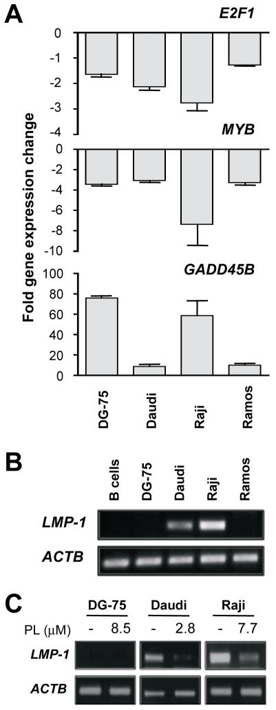
Figure 5. Treatment with PL changes expression of cellular NF-κB/MYC target genes and the EBV-encoded oncogene, LMP-1.
(A) BL cells treated with PL at IC50 for 24 hrs exhibit reduced expression of E2F1 and MYB and increased expression of GADD45B. Total RNA was analyzed using qPCR. Gene expression levels were normalized to levels of HPRT message and expressed as fold change in PL-treated cells versus untreated cells. Mean values and standard deviations of an experiment performed in triplicate are presented as columns and error bars, respectively.
(B) RT-PCR detects LMP-1 mRNA in EBV+ Daudi and Raji cells but not in EBV− BL (DG-75 and Ramos) or normal peripheral blood B cells.
(C) PL inhibits LMP-1 expression in Daudi and Raji cells. DG-75 was included as control. BL cells (1 × 10⁶/ml) were treated with PL at IC50 for 24 hrs. Total RNA was prepared and analyzed using RT-PCR.

