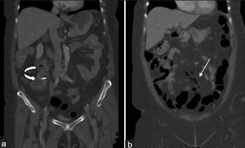
Figure 1.

(a and b) Contrast-enhanced coronal CT images (lung window) demonstrate linear pockets of air tracking within small mesenteric veins (white arrow). Note the enteroscopically placed clip in the patient's distal ileal GIST, which was tattooed for the surgeon (curved arrow).
