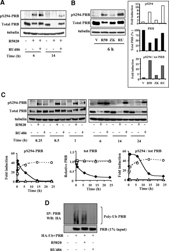
Fig. 2.

Unlike R5020 and ZK98299, RU486 induces stable PRB- pS294. A, Ishikawa PRB cells were treated by ligands as in Fig. 1 during 6 or 14 h, and whole cells extracts were immunoblotted using PRB serine 294 phospho-specific antibody, anti-PRB, or antitubulin antibody. B, Ishikawa PRB cells were treated with vehicle (V) or R5020 (R50) (10−8 m) or ZK98299 (ZK) (10−6 m) or RU486 (RU) (10−8 m) during 6 h, and whole cell extracts were immunoblotted as in A. Numerized band densities corresponding to pS294-PRB (upper inset) or total PRB (middle inset) are normalized to vehicle or tubulin controls and plotted as fold induction or percentage of total PRB in ligand-free condition. Ligand-induced pS294/PRB (lower inset) is presented as fold induction of vehicle-treated cells. C, Ishikawa PRB cells were treated without or with R5020 (10−8 m) or RU486 (10−8 m) during the indicated time periods, and whole cell extracts were immunoblotted as in A. pS294-PRB and PRB band densities were normalized to vehicle or tubulin controls and plotted as fold induction of ligand-free species for each time point (left and middle panels), and the corresponding ratio is shown in the right panel (white triangle, vehicle; black diamond, R5020; and white circle, RU486). D, Parental Ishikawa cells lacking PRB expression were transiently transfected with HA-ubiquitin and PRB expression vectors during 48 h, pretreated with MG132 (5 μm) during 30 min, and then incubated without or with R5020 (10−8 m) or RU486 (10−8 m) during 4 h. After PRB immunoprecipitation (IP) using monoclonal anti-PR antibody, ubiquitinated-PRB was analyzed by Western blotting (WB) using anti-HA antibody (upper panel). PRB levels corresponding to 1% input were detected by anti-PR antibody (lower panel).
