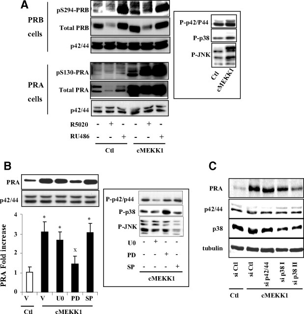
Fig. 7.
MEKK1-induced PRA stabilization is impaired by p38 inhibition. A, Ishikawa PRB or PRA cells were transiently transfected with either empty control (Ctl) vector or cMEKK1 expression vector during 24 h and then treated without or with R5020 (10−8 m) or RU486 (10−8 m) during 24 h. Whole cell extracts were immunoblotted, and P-p42/44, P-p38, and P-JNK MAPK levels were detected by specific antibodies (inset). B, Ishikawa PRA cells were pretreated with vehicle (V) or U0126 (U0) (10 μm) or PD169316 (PD) (10 μm) or SP600125 (SP) (10 μm) during 30 min and then transfected with empty or MEKK1 expression vector during 24 h. Immunoblot analysis was performed as above, and normalized PRA band intensities are presented as fold increase as compared with PRA levels in control cells (inset). Statistical significance is represented by asterisks when comparison is done between control or cMEKK1 condition and by XX when selective MAPK inhibition is compared with nontreated MEKK1-transfected cells. C, Ishikawa PRA cells were cotransfected with control or cMEKK1 vector along with either control or specific siRNA against both p42 and p44 or p38 MAPK during 24 h as described in Materials and Methods. Cells were then incubated in 5% fetal calf serum containing medium for another 48 h before performing immunoblot analysis.

