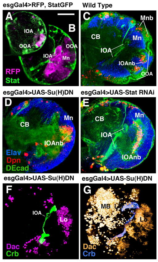
Figure 6.
Optic lobe-directed expression of mutant constructs using the esg-Gal4 driver line. (A, B): Frontal confocal sections of first instar (A) and early third instar (B) brain showing RFP expression, driven by esg-Gal4, in optic lobe (magenta). Stat92EGFP is labeled in green. (C) Frontal confocal section of third instar wild-type brain labeled with anti-DEcad (green), anti-Elav (blue), and anti-Deadpan (red). CB, central brain; IOA, inner optic anlage; IOAnb, neuroblasts of inner optic anlage; Mn, medulla neurons; Mnb, medulla neuroblasts; OOA, outer optic anlage. (D, E): Frontal confocal sections of late third instar brain in which a dominant-negative Su(H) construct [Su(H)DN] (D) or Stat92E-RNAi construct (E) under UAS control was driven by esg-Gal4. Note loss of epithelial outer optic anlage and premature growth of medullary neurons. (F, G) Labeling with anti-Dachshund (Dac) illustrates strong reduction of lamina primordium (La) in confocal section (F) and volume rendering (G). MB, mushroom body (central brain).
Bar: 40μm

