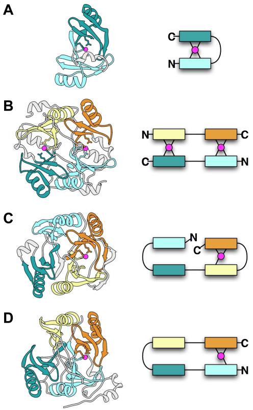
Figure 2.
Examples of alternate arrangements of paired beta-alpha-beta-beta-beta modules in the VOC SF. Structures are shown on the left and schematic diagrams on the right. In both the structures and schematics, metal ions are magenta and different modules are shown in different colors to highlight the repeating beta-alpha-beta-beta-beta unit. Metal-ligating residues may occur in the first and/or last beta-strands of a module. Parts of the structures not within beta-alpha-beta-beta-beta modules are shown in light gray. (A) Glyoxalase I from Clostridium (PDB 3HDP), in which two modules from a single chain pair to form a metal site. (B) Human glyoxalase I (PDB 1QIN), in which two chains each containing two modules pair in head-to-tail fashion to form two metal sites. (C) 2,3-Dihydroxybiphenyl 1,2-dioxygenase from Burkholderia (PDB 1KMY), in which the four modules in a single chain pair in the order 1-2 and 3-4, and only the latter pair forms a metal site. (D) Protein of unknown function from Bacillus (PDB 1ZSW), in which the four modules in a single chain pair in the order 1-4 and 2-3, and only the former pair forms a metal site.

