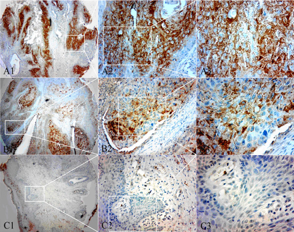
Figure 3.
Expression pattern of αB-crystallin in tumor tissue and tumor-adjacent tissue of LSCC. TMA sections were analyzed by immunohistochemical staining. Brown staining indicated positive expression of αB-crystallin. A1-3: The expression pattern of αB-crystallin in moderately differentiated LSCC tissue. B1-3: The expression pattern of αB-crystallin in well-differentiated LSCC tissue. C1-2: The expression pattern of αB-crystallin in tumor-adjacent tissue with weakly positive staining of αB-crystallin. C3: Squamous epithelium of adjacent nontumorous tissue with negative staining of αB-crystallin. Original magnification: ×40 in A1, B1 and C1; ×100 in A2, B2 and C2; ×400 in A3, B3 and C3.

