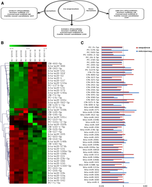
Figure 5.

Differential miRNA expression in the mammary gland. (A) The Venn diagram displays the distribution of 884 unique miRNAs found in lactation and non-lactation periods. (B) The heat map was constructed based on the mean expression levels of the microarray. (C) Comparison between the microarray (blue bar) and sequencing (red bar) data. The bar graph shows the ratio of lactation to non-lactation (R value), where R values of higher or lower than 1 in both methods indicate that the miRNA has the same expression trend in the two different assay systems. R values where R(microarray)>1 and R(sequencing)<1 indicate that miRNA changes are differently represented by the two methods.
