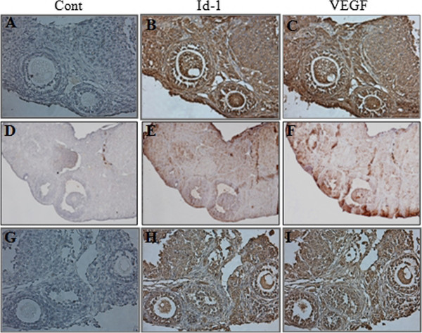
Figure 2.
Immunohistochemical analysis of Id-1 and VEGF in ovaries. Whole ovaries were collected 18 hours after hCG injection. (A-C), young control mice; (D-F), aged control mice; (G-I), 0.1 ng BMP-6-treated aged mice. Control was immunostained without primary antibody (purple color) (A, D, G). Cell immunostained with Id-1 (B, E, H) and VEGF (C, F, I)-specific antibody showed a brown color. n =3, six ovaries per aged group (x 100 magnifications).

