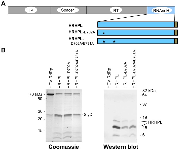
Figure 4. Recombinant HBV RNAseH proteins.
A. Structure of the recombinant RNAseHs. The HBV polymerase with its major domains labeled is at top. The recombinant RNAseH derivatives are shown below with the C-terminal hexahistidine tag in brown. TP, terminal protein domain; RT, reverse transcriptase domain; *, mutations D702A or E731A to RNAseH active site residues. B. Proteins in the enriched lysates. The left panel is a Coomassie-blue stained SDS-PAGE gel of enriched RNAseH extracts as employed in the RNAseH assays. The right panel is a western blot of the extracts employing monoclonal antibody 9F9 which recognizes an epitope near the C-terminus of the HBV polymerase.

