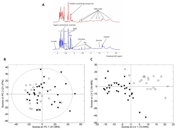
Figure 1.
Umbilical cord blood profile. Superposed 1 H NMR spectra for the spectra of umbilical cord blood plasma (Panel A), Principal Component Analysis scores plot (PCA, panel B), and Projection to Latent Structures for Discriminant Analysis scores plot (PLS-DA, panel C) showing the global metabolic differences between low birth weight (open circles, blue spectra) and control birth weight (black triangles and red spectra) newborns. Although an unsupervised analysis by PCA does not show differences between groups, a supervised classification method (PLS-DA) provides a differential global metabolic profile. The cross-validated error percentage of the model for the classification of low weight at birth samples is 8%. Metabolites exhibiting largest differences between groups have been listed in Table 2.

