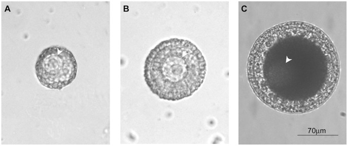
Figure 1. Representative images of the three GV stages in oocytes of Styela plicata.
Panels A), B) and C) represent pre-vitellogenesis (stage A; GV-A), vitellogenesis (stage B; GV-B) and post-vitellogenesis (stage C; GV-C), respectively. The germinal vesicle is indicated (arrow head) in all stages reproduced.

