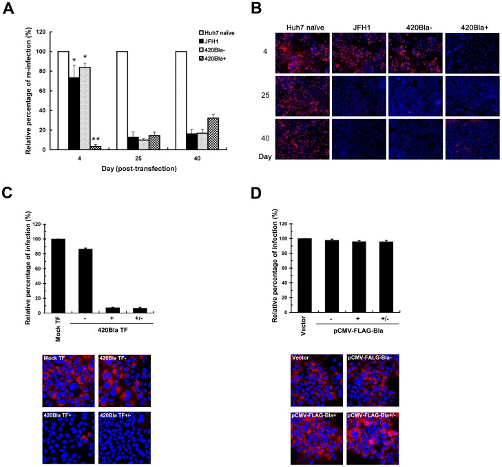
Figure 6. Interference with HCV reinfection in 420Bla+ stable cells as well as in chronically infected cells.
(A and B) Huh7 cells were transfected with JFH1 or 420Bla RNAs, and cultured in the absence or presence of blasticidin. Huh7 naïve cells indicate the mock-transfected cells cultured in parallel. At the indicated times, Huh7 naïve, and JFH1-transfected 420Bla− and 420Bla+ cells were infected with 420RFP virus at an MOI of 0.05 for 12 hr and cultured for additional 3 days. Half of the 420RFP infected cell samples was quantified by flow cytometry (A) and immunofluorescence microscopy (B), respectively, for RFP-positive cells. The relative percentage of reinfection in viral RNA transfected cells was calculated by normalization with that of Huh7 naïve cells in (A). The Hoechst 33258 staining (blue) in (B) indicates the nuclei. (C) The Mock TF, 420Bla−, 420Bla+, and 420Bla+/− cells were established as described in Figure 3A, and the cells were analyzed for 420RFP reinfection as described above. (D) The Vector, FLAG-Bla−, FLAG-Bla+, and FLAG-Bla+/− cells from Figure 3E were also assayed for 420RFP virus reinfection as described above. Data represents mean ± SEM (n = 3).

