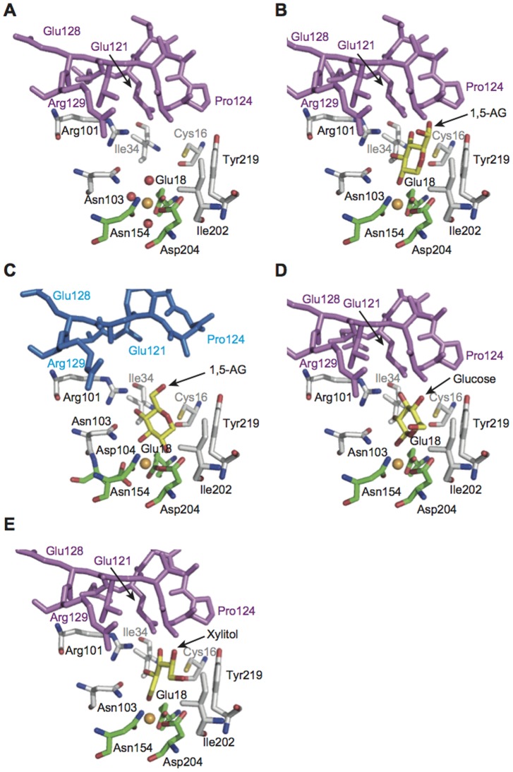
Figure 4. Active site structures of mouse and human SMP30/GNL.
(A) Mouse SMP30/GNL in the substrate-free form, (B) the mouse SMP30/GNL–1,5-AG complex, (C) the human SMP30/GNL–1,5-AG complex, (D) the mouse SMP30/GNL–d-glucose complex, and (E) the mouse SMP30/GNL–xylitol complex. Lid loop residues of mouse SMP30/GNL and human SMP30/GNL are shown in purple and blue, respectively. Carbon atoms of ligand residues for the divalent metal ion (orange sphere) and those for substrate/product analogues are shown in green and yellow, respectively. Other carbon atoms are shown in white.

