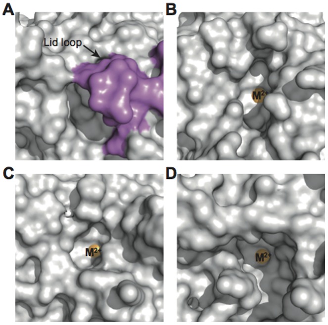
Figure 5. Top view of the substrate-binding cavity.

Surface representation of (A) mouse SMP30/GNL, (B) DFPase, (C) Drp35, and (D) PON. Residues in the lid loop of mouse SMP30/GNL and the divalent metal ions (labeled as M2+) are shown in purple and orange, respectively. Structures of DFPase, Drp35, and PON are superposed onto mouse SMP30/GNL by the SSM fitting using the program Superpose [27] in the CCP4 program suite [20], and all molecules are viewed from the same direction.
