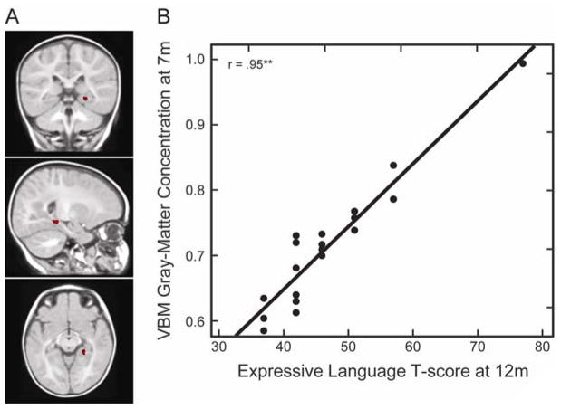
Figure 4.
(A). Coronal (y = −25), sagittal (x = 19), and axial (z = −9) view of the right hippocampus at 7 months. (B) VBM gray-matter concentration at 7 months, measured as cubic millimeters of gray-matter per voxel in the right hippocampus region, as a function of expressive language skills at 12 months.

