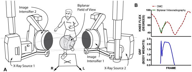
Figure 1.
A, experimental set-up including image intensifiers and x-ray sources. The optical motion capture cameras are not shown. The subject is performing the jump-cut maneuver. In this example they were cued to cut to their left upon landing on the force plate. The ‘X’ marks the landing location and the arrows represent the left (L) and right (R) cut directions. B, the OMC (dotted red) and biplanar videoradiography (solid green) knee flexion/extension and GRF (solid blue) for the entire jump-cut activity including the flight phase, landing, rotation, cut, and toe-off. The field of view for the biplanar videoradiography limits its ability to collect kinematic data for the entire jump-cut activity. However, it can be tailored to measure motion for specific periods of an activity where OMC is more sensitive to soft tissue artifact.

