
Fig. 2.
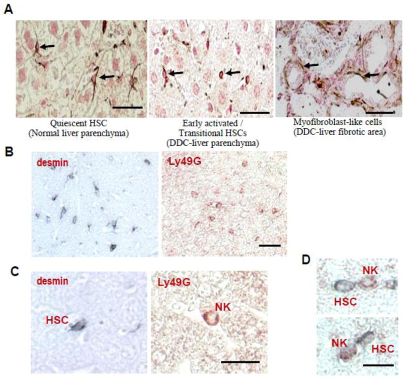
In vivo evidence for the contact between NK cells and early activated HSCs. (A): Identification of early activated/transitional HSCs and NK cells in the fibrotic livers from DDC-fed mice. Under the light microscope, early activated desmin-positive cells (arrows in the middle panel) appear as small cells with scant cytoplasm and lack numerous processes typical for quiescent HSCs (arrows in the left panel). Bars, 50 μm. (B): Mice were fed a DDC diet for 2 weeks. Liver sections were stained with desmin and Ly49G antibodies to identify HSCs and NK cells, respectively. Positive staining was developed using either 3,3′-diaminobenzidine containing nickel chloride (black staining for HSCs) or 3-amino-9-ethylcarbazole (red staining for NK cells). Note that positive cells occupied a similar area of the liver parenchyma, including zones 2 and 3. (C): Higher magnification of HSCs and NK cells located in the perisinusoidal space of Disse and in the hepatic sinusoid, respectively. (D): Double immunostaining of NK cells and HSCs demonstrating that Ly49G-positive NK cells (red) are located in close proximity to desmin-positive (black) early activated HSCs. Bars, 50 μm.