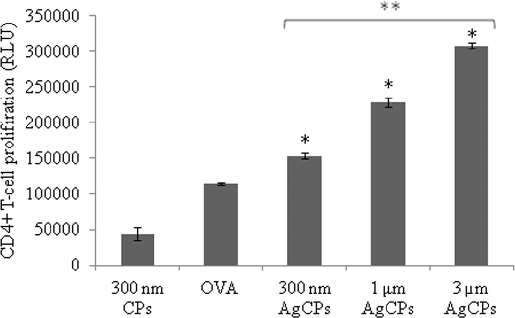
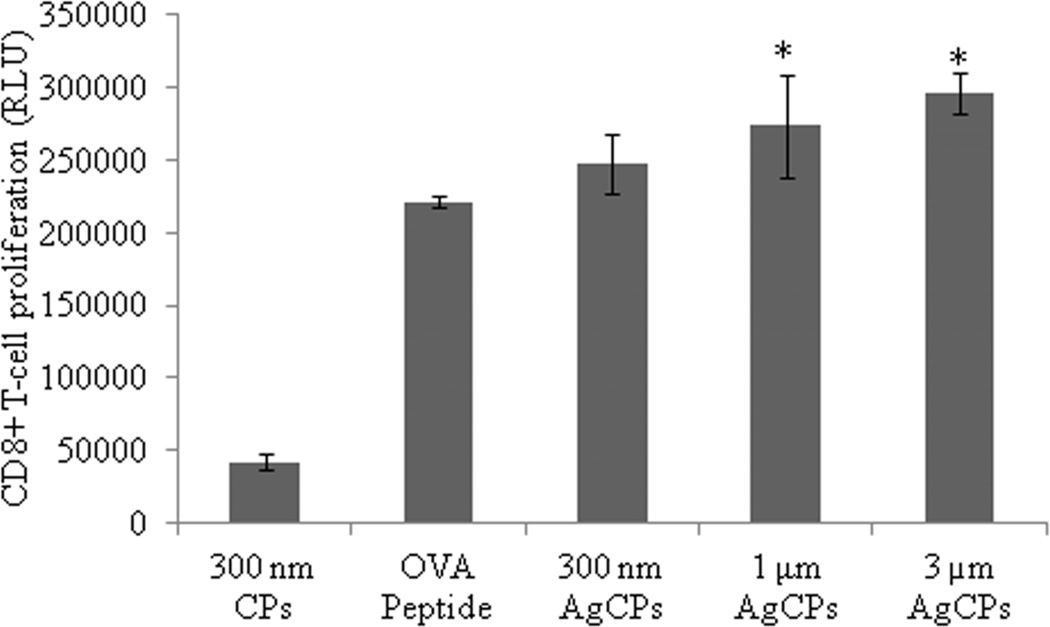
Fig. 7.
Proliferation of (a) OVA-specific CD4+ T-cells, and (b) OVA-specific CD8+ T-cells in response to presentation of antigen by BMDCs. BMDCs were pulsed with CPs, AgCPs, soluble full length OVA or the MHC I-restricted OVA257–264 peptide for 24 hrs and then co-incubated with CD4+ or CD8+ T-cells from OT-II or OT-I mice, respectively. After 72 hrs, T cell proliferation was determined via a non-radioactive proliferation assay (CellTiter Glo; Promega, Madison, WI). Data are presented as mean relative light unit (RLU) ± standard deviation from one of two independent experiments with similar results. *p ≤ 0.05 vs. OVA or OVA peptide; **p ≤ 0.05 vs other AgCP sizes.


