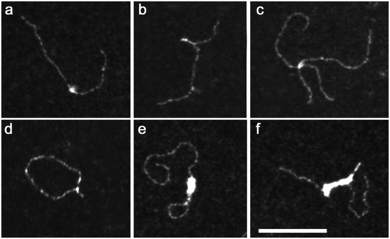
Figure 4. Complexes formed between IN and dsDNA observed by TEM.
Reactions were carried out for 10 min. in 10mM HEPES (pH 7.5), 100mM NaCl, 7.5mM MgCl2 using 3.8nM of a HIV-1 derived DNA fragment (800bp) and 125nM IN monomer (nucleotide/IN ratio: 32). The most representative individual IN-DNA complexes have been selected once formed at 37°C (a-d) or at 4°C (e-f). The scale bar is the same for all panels and represents 100 nm.

