Figure 4.
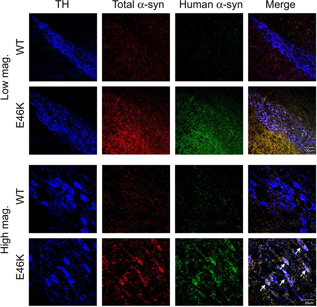
α-Synuclein accumulation and aggregation in the substantia nigra. Animals (12-month old) expressing E46K-mutated α-synuclein exhibit overt accumulation and aggregation of α-synuclein. Low magnification images show diffuse accumulation of both total and aggregated human α-synuclein. High magnification images show both neuropil (likely in processes) and intracellular accumulation and aggregation in nigral dopamine neurons (tyrosine hydroxylase +, TH). The morphology of E46K neurons is also altered compared to wild-type (WT; littermate controls), where the cell membrane appears to be undergoing fragmentation (indicated by arrows).

