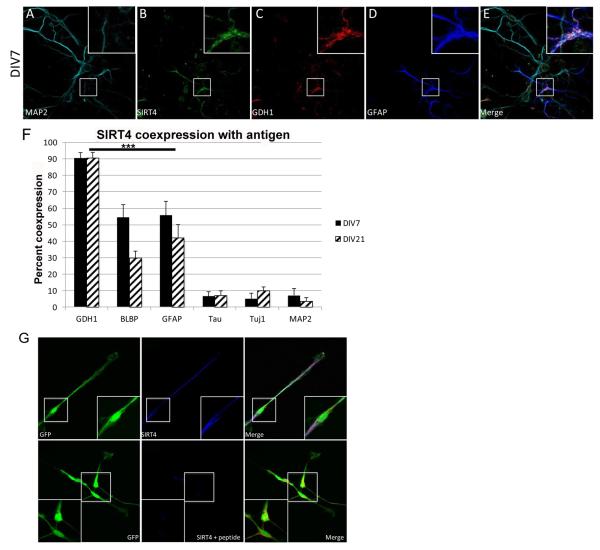
Figure 2. Expression of SIRT4 and GDH1 in dissociated cortical cultures.
A-D: Expression of the indicated protein in mixed neuronal/glial co-cultures from E18 cortical cultures, shown at DIV7, 60X magnification. E: Merged image of A-D. Boxed area in center indicates additional 3.4X magnified area at top right of each image (2A-E and 2G). F: Quantitation of percentage of co-expression of SIRT4 with indicated protein at DIV7 and DIV21. ***p< 0.001. GDH1, BLBP and GFAP are significantly different from tau, Tuj1 and DAPI staining. BLBP does not differ from GFAP. ***p < 0.001. Co-expression with GDH1, BLBP and GFAP is significantly different than co-expression with tau, Tuj1 and DAPI. G: Expression of GFP in CTX cells at DIV2. SIRT4 expression without and with SIRT4 specific antigenic peptide preincubation.

