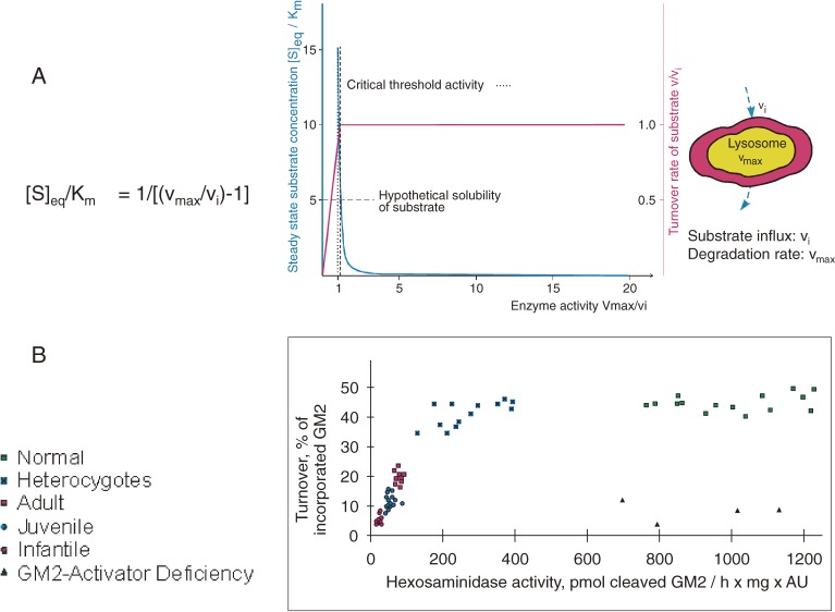
Figure 4.
Residual catabolic activity correlates with clinical forms of GM2 gangliosidoses (modified after ref. 53). A) Steady-state substrate concentration as a function of enzyme concentration and activity.51) The model underlying this theoretical calculation assumes influx of the substrate into the lysosomal compartment at a constant rate (vi) and its subsequent utilization by the catabolic enzyme (for details see text). Blue line = [S]eq steady-state substrate concentration; ……… = theoretical threshold of enzyme activity; - - - - - - - = critical threshold value, taking limited solubility of substrate into account; red line = turnover rate of substrate (flux rate). B) In order to experimentally verify the basic assumptions on which this model rests, studies were performed in cell culture.53) The radiolabeled substrate ganglioside GM2 was added to cultures of skin fibroblasts with different activities of beta-hexosaminidase A and its uptake and turnover measured. The correlation between residual enzyme activity and the turnover rate of the substrate was essentially as predicted: degradation rate of ganglioside GM2 increased steeply with residual activity, to reach the control level at a residual activity of approximately 10–15% of normal. All cells with an activity above this critical threshold had a normal turnover. Comparison of the results of these feeding studies with the clinical status of the donor of each cell line basically confirmed our notions but also revealed the limitations of the cell culture approach.

