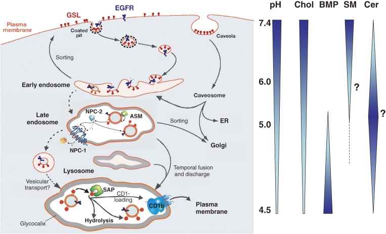
Figure 8.
Principles of lysosomal sphingolipid catabolism and membrane digestion. Proposed topology of endocytosis and lysosomal degradation.130) A section of the plasma membrane is internalized by way of coated pits or caveolae. These membrane patches include glycosphingolipids (GSL, red) and receptors such as EGFR (epidermal growth factor receptor, blue). These vesicles fuse with the early endosomes which mature to late endosomes. Endosomal perimeter membranes form invaginations, controlled by ESCRT proteins,129) which bud off, forming intra-endosomal vesicles. Lipid sorting occurs at this stage. The pH of the lumen is at about 5. At this pH, acid sphingomyelinase is active and degrades sphingomyelin of the intra-endosomal vesicles to ceramide, whereas the perimeter membrane is protected against the action of ASM by the glycocalix facing the lumen. This reduction of the sphingomyelin level, coupled with the increase in ceramide, facilitates the binding of cholesterol to NPC-2 and its transport to the perimeter membrane of the late endosome where it is transferred to NPC-1.261) This protein enables the export of cholesterol through the glycocalix, eventually reaching cholesterol binding proteins in the cytosol. Ultimately, late endosomes fuse with lysosomes. The GSLs are in intra-lysosomal vesicles facing the lumen of the lysosome and are degraded by hydrolases with the assistance of LLBPs. The products of this degradation are exported to the cytosol or loaded on CD1b immunoreceptors and exported to the plasma membrane for antigen presentation. Gradients of pH in the lysosol, and intra-endo-lysosomal vesicle content of cholesterol (Chol), BMP, sphingomyelin (SM, hypothetical) and ceramide (Cer; hypothetical) are shown (modified after ref. 83).

