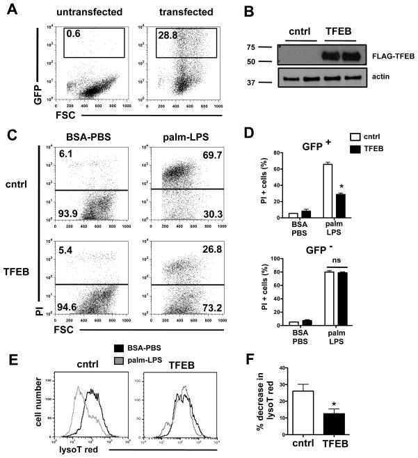
Figure 7. Over-expression of TFEB prevents lysosomal phenotype and rescues cell death induced by palmitate and LPS.
HEK293-TLR4/CD14/MD2 cells were transfected control GFP vector (cntrl) or a FLAG-TFEB expression construct containing an IRES-GFP element. Cells were stimulated as indicated 24 h after transfection and flow cytomery was performed 48 h later. (A) Approximately 29% of the total cells expressed the GFP construct. (B) TFEB protein expression was assessed by western blot 24 h after transfection using α-FLAG antibody. Actin is shown as loading control. (C, D) 24 h following transfection, cells were stimulated with BSA-PBS or palm-LPS for 48 h, stained with PI and analyzed by flow cytometry. Representative dot plots from the GFP+ cells are shown (C), and summed data for GFP+ (top panel) and GFP− (bottom panel) cells are plotted (D). (E) LysoT red fluorescence was assessed 48 h after stimulation with BSA-PBS (black) or palm-LPS (gray) in control vector and TFEB-transfected cells. Representative histograms are shown. (F) Quantification of the palm-LPS induced decrease in lysoT red fluorescence (MFI) in control and TFEB transfected cells. All experiments were performed a minimum of 3 times in triplicate. Bars represent mean ± SE. *, p ≤ 0.05 for control vector vs. TFEB vector; ns, non-significant.

