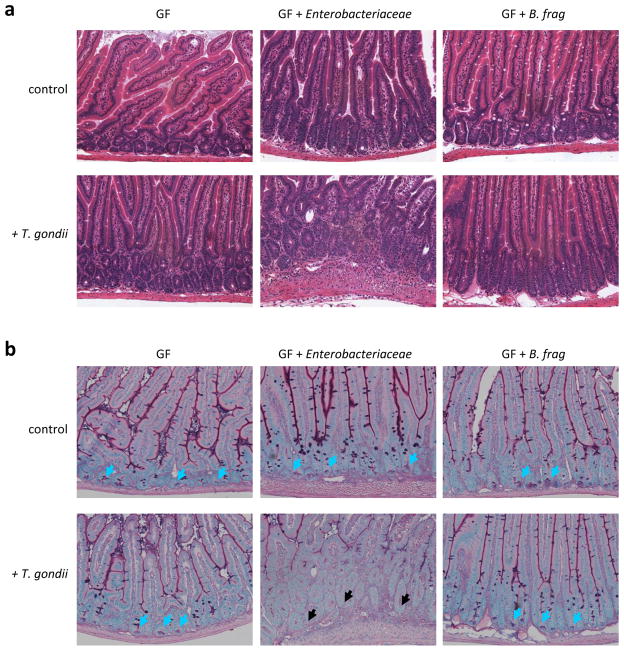
Figure 3. T. gondii-induced dysbiosis contributes to the intestinal pathology.
(a) Germ-free (GF) mice were left untreated, colonized with the Enterobactericeae isolate (Supplemental Fig. 3a), or with Bacteroides fragiles (B. frag), and infected orally with 20 cysts of the T. gondii ME49 strain. Histological analysis of the small intestine was performed on day 7 post-infection. The results are representative of two independent experiments. (b) Germ-free (GF) mice were mock colonized, colonized with the Enterobactericeae isolate (Supplemental Fig. 3a), or with B. frag, and additionally infected orally with T. gondii. Histological visualization of Paneth cells in the small intestines were performed on day 7 post-infection. The blue arrows indicate Paneth cells and the black arrows point toward bases of the crypts lacking these cells. The results are representative of two independent experiments.

