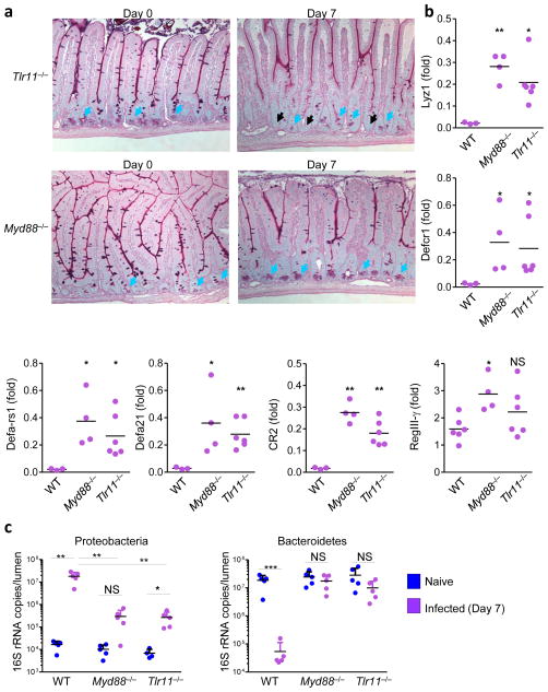
Figure 4. TLR11-mediated activation of MyD88 triggers Paneth cell death and intestinal dysbiosis.
(a) WT (not shown), Tlr11−/−, and Myd88−/− mice (five mice per group) were left untreated or were infected orally with 20 cysts per mouse of the ME49 strain of T. gondii. Histological visualization of Paneth cells in the small intestines were performed on day 7 post-infection. The blue arrows indicate Paneth cells and the black arrows point toward bases of the crypts lacking these cells. (b) The relative expression levels of Defcr1 (alpha-defensin-1), Defa- rs1, Defa21, CR2, and RegIII-γ were measured by qRT-PCR in the small intestines of WT (n=3), Tlr11−/− (n=6), and Myd88−/− (n=4) mice on day 7 post-infection. * P< 0.05; ** P< 0.01. (c) qRT-PCR analysis of Proteobacteria and Bacteroidetes in the lumens of small intestines of naïve (blue) or T. gondii-infected (purple) mice. The data shown are the means ± SD. * P< 0.05; ** P< 0.01; *** P< 0.001. The results are representative of >10 independent experiments, each involving 3–7 mice per group.

