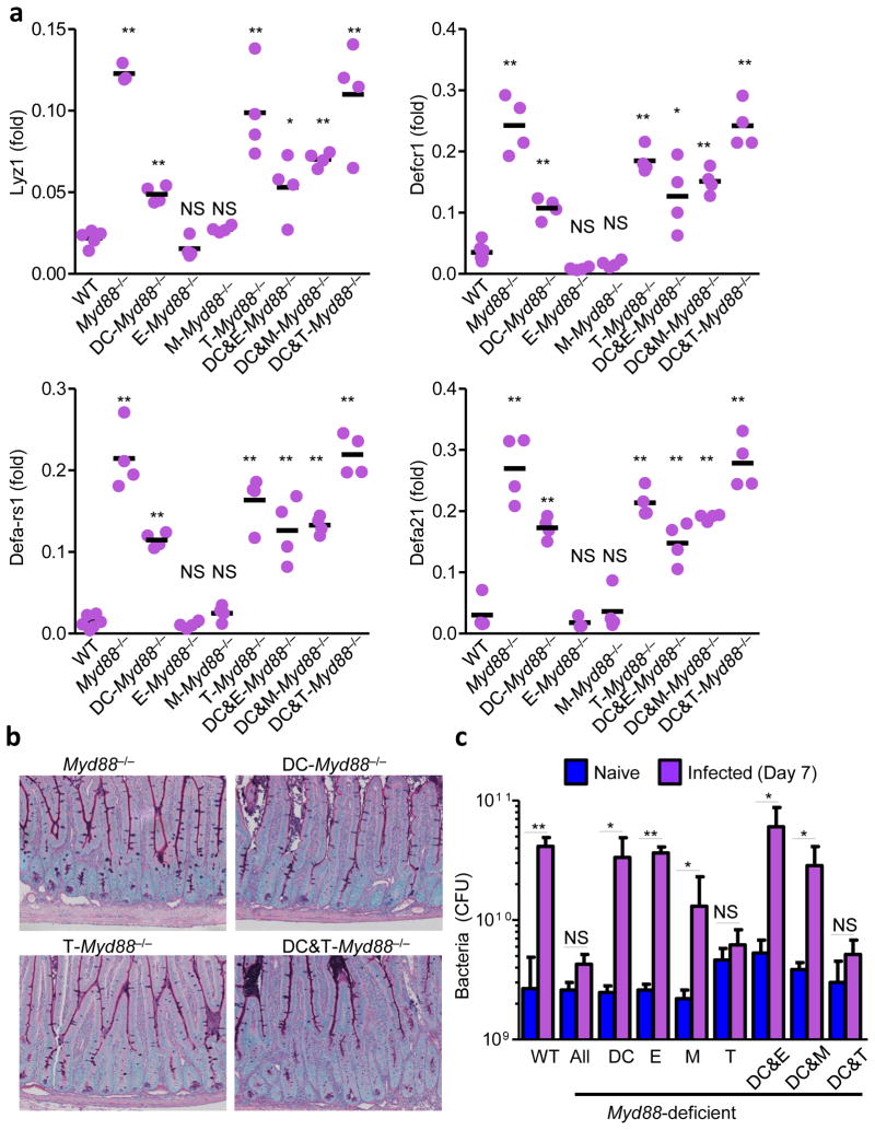
Figure 7. Intrinsic T-cell MyD88 signaling mediates loss of Paneth cells intestinal dysbiosis.
(a) qRT-PCR analysis of relative expression of Lyz1, Defcr1 (alpha-defensin-1), Defa-rs1, Defa21 in the small intestines of WT, Myd88−/−, and cell type-specific MyD88-deficient mice on day 7 post-infection. The results are representative of five independent experiments each involving 4–5 mice per group. (b) Histological visualization of Paneth cells in small intestines of DC-Myd88−/−, T-Myd88−/−, and DC&T-Myd88−/− mice compared to complete MyD88-deficient mice (Myd88−/−). The results are representative of five independent experiments each involving 4–5 mice per group. (c) Bacterial loads in the lumens of small intestines of naïve (blue) or infected WT, Myd88−/− and cell type-specific Myd88−/− mice (purple) were analyzed by plating bacteria on blood agar plates on day 7 post-infection. * P< 0.05; ** P< 0.01. The results are representative of five independent experiments each involving 4–5 mice per group.

