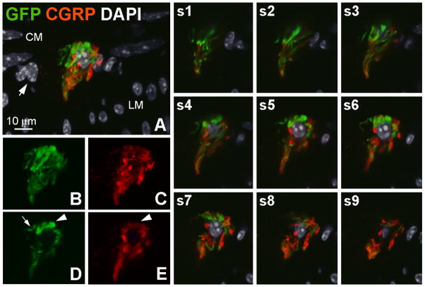
Figure 5.
A globular cluster of GFP-ir and CGRP-ir fibers. A, GFP- (green) and GCRP-positive (red) nerve fibers surrounding a nucleus labeled by DAPI (gray). DAPI-labeled small oval nuclei and elongated nuclei delineate the longitudinal and circular muscle layers, respectively. Nuclei located between these two layers (arrow), including the nucleus surrounded by nerve fibers, likely belong to neurons within a myenteric ganglion. The image is a projection of 9 slices 1 μm apart. B–E, Clusters of GFP- and CGRP-positive fibers are shown in B and C, respectively, (9 slices) and in a single slice (D and E). Arrowheads in D and E indicate a structure that is intensely GFP-positive and weakly CGRP-positive. The arrow in D indicates a segement of a fiber that is GFP-positive and CGRP-negative. Images s1–s9 show the relationship of GFP- and CGRP-ir fibers to the nucleus in the center of the cluster in individual optical sections. Scale bar: 10 μm.

