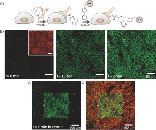
Figure 3.
Spatiotemporally controlled dual-color labeling of the A431 cells. A) Antibody mediated two-step labeling approach. TCO-Ab against the biomarker of interest (EGFR) were targeted to A431 cells and then used as scaffolds for bioorthogonal coupling of Tz-PANP in live cells. B) Left inset, a CLSM image of the A431 cells showing VT680 fluorescence (pseudo-colored red) from the cell surface. Left, unlike the VT680 channel, no fluorescence signal was detectable in the fluorescein channel. Center and right, subsequent exposure of the cells to 405 nm laser light resulted in fluorescein fluorescence (pseudo-colored green) from the cell surface. Fluorescence intensity increased with increasing light exposure time. C) A CLSM image showing the presence of fluorescein fluorescence from the cell surface, restricted to the photoactivated region of the imaging slide (central square). D) A merged CLSM image showing fluorescent signals from both the fluorescein and VT680 channels. The light exposed area (central square) contains dual-color labelled A431 cells.

