
Figure 1.
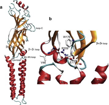
(a) Structural model of the α-subunit from the Torpedo AChR (PDB code 2BG9). (b) Close-up view of the region between the ligand-binding and pore domains. Key residues are highlighted in stick representation and colored according to electronic charge (blue, positive; red, negative; and gray, neutral).