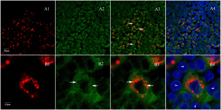
Figure 2. Relationship of labeled sympathetic nerve endings and innervated cells.
A plasma membrane dye, DiO, was used to stain the histological section of the lymph nodes labeled with FR and was then examined with confocal microscopy (TCS SP5, Lecia). Micrographs showed that FR-labeled nerve endings (red) landed on the surface of certain cells in lymph nodes (A, arrows). These cells are large in size, mononucleated with light nuclear density (DAPI staining). (B) Nerve endings wrapped around the targeted cell (arrows in B2 and B3) on an enlarged scale. The innervated cells were surrounded by lymphocytes (arrow heads in B4). A1 and B1, FR-labeled sympathetic nerve fibers endings; A2 and B2, Dio-labeled cell membranes; A3 and B3, merged images of FR and Dio from A1-A2 and B1-B2, respectively; A4 and B4, merged images of A1-3 and B1-3, respectively.

