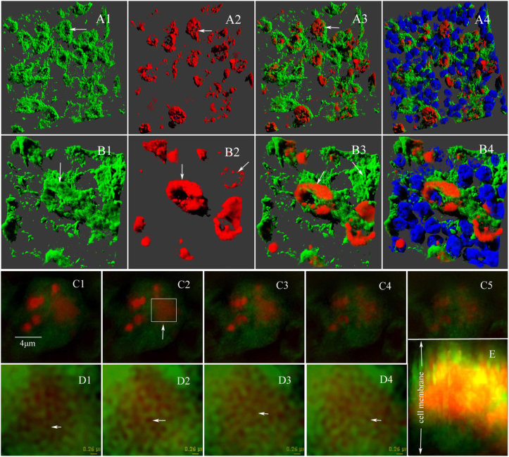
Figure 3. Three-dimensional reconstruction and high-resolution SIM observation of labeled-nerve endings and innervated cells.
(A, B) 3D reconstruction clearly showed that the labeled nerve endings (red) land on the cell membrane (green) of innervated cells (B, high magnification). (C) A series of single-cells images (0.4 μm) acquired by Z-stack scanning from high-resolution SIM (Total internal reflection fluorescence 100×1.49 NA oil immersion, Nikon) revealed a mosaic structure of nerve endings with the membrane of the innervated cell. (D) Enlargement of the image in the area indicated in C2. The enlargedimages clearly showed 0.1–0.2 μm of labeled nerve endings in the cell membrane of the innervated cell. (E) 3D reconstruction of a series of images (0.4 μm) acquired by Z-stack scanning with high-resolution SIM.

