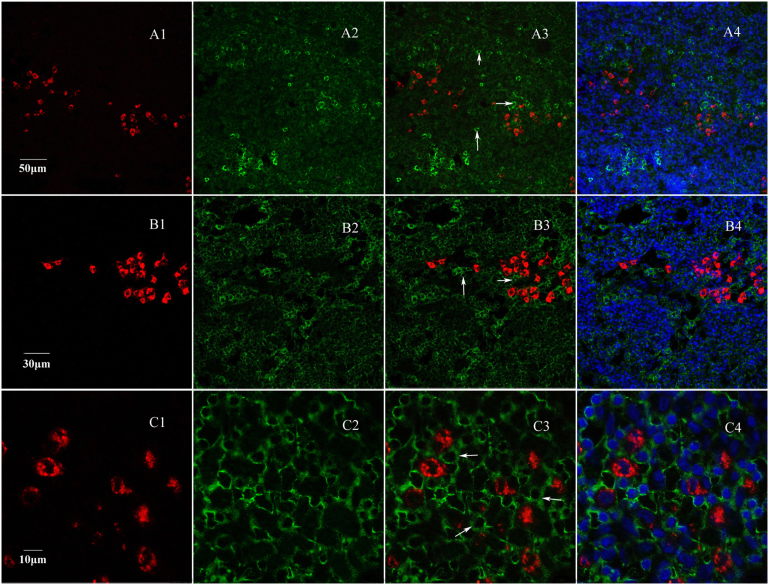
Figure 4. Fluorescence immunocytochemistry for CD3, CD22 and CD14 with slice labeled by FR.
Red fluorescence (A1,B1,C1) is labeled nerve fibers, and green fluorescence (A2,B2,C2) is CD3+,CD22+and CD14+cells. A3, B3 and C3 are respectively merged images from CD3+ ,CD22+ and CD14+ cells and FR-labeled nerve fibers. The merged images showed that labeled nerve fibers were not around CD3+, CD22+ and CD14+ cells (A3, B3, C3 and A4, B4, C4). This immunofluorescence results demonstrated that the nerve fibers from superior cervical ganglion do not target T cells, B cells and macrophages.

