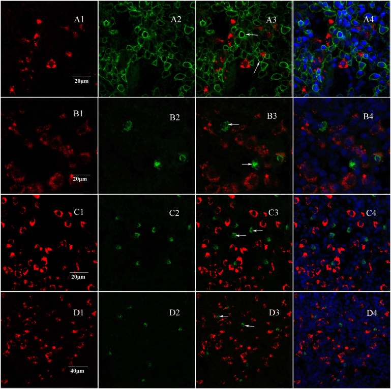
Figure 5. Fluorescence immunocytochemistry for MHCII, CD11c, CD103 and CD138 with slice labeled by FR.
Red fluorescence (A1, B1, C1, D1) is labeled nerve fibers, and green fluorescence (A2, B2, C2, D2) is MHCII+, CD11c+, CD103+ and CD138+ cells. A3, B3, C3 and D3 are respectively merged images from MHCII+, CD11c+, CD103+ and CD138+ cells and FR-labeled nerve fibers. The merged images showed that labeled nerve fibers were not around MHCII+, CD11c+, CD103+ and CD138+ cells (A3, B3, C3, D3 and A4, B4, C4, D4). This immunofluorescence results demonstrated that the nerve fibers from superior cervical ganglion also do not target dendritic cells and plasma cells.

