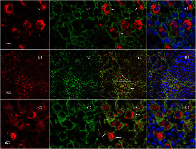
Figure 6. Fluorescence immunocytochemistry of S100 protein and SYP protein.
(A) Immunocytochemistry of S100 showed that cells targeted by FR-labeled nerve endings were S100 protein positive (green, arrows). (B) SYP staining showed that FR-labeled nerve endings were completely overlapped with SYP proteins (green, arrows), indicating that labeled nerve endings expressed SYP proteins. (C) Enlarged images in the area indicated in B4. A1, B1 and C1, FR labeled sympathetic nerve fibers; A2, S100 staining; B2 and C2, SYP staining; A3, B3 and C3, merged images of A1-2, B1-2 and C1-2, respectively; A4, B4 and C4, merged images of A1-2-3, B1-2-3 and C1-2-3, respectively.

