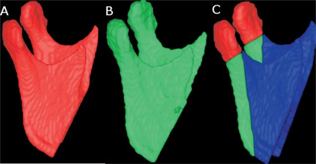
Fig 2.
Lateral views of 3D models of patient. A, 3D model constructed from CBCT image acquired 1-2 weeks before surgery. B, 3D model labeled green constructed from CBCT scan 1 week postsurgery. Other anatomic structures are masked for better visualization of changes in mandibular ramus and condyle. C, A and B are combined after superimposition to identify regions of interest in mandibular rami: condyles (red) and posterior border (green).

