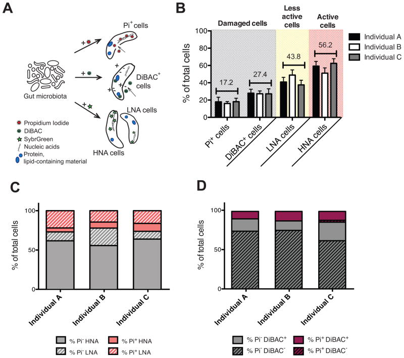
Figure 1. The human gut microbiota is highly active with substantial proportions of damaged cells.
Also see Figures S1, S2 and Table S3. (A) Cellular targets of the fluorescent dyes. The nucleic acid dye Pi enters cells with compromised membranes; DiBAC binds to intracellular lipid-containing material of depolarized cells; and SybrGreen stains the nucleic acids of all bacteria irrespective of their membrane status, identifying two clusters of cells: the low- (LNA) and high- (HNA) nucleic acid containing cells. (B) Average proportions of damaged cells (Pi+ and DiBAC+) and cells with a low (LNA) or high nucleic acid content (HNA) in three unrelated individuals (n=5–10 samples/individual). Values are mean±sem. (C) SybrGreen and Pi dual-staining in 3 unrelated individuals. Pi- cells are in grey, Pi+ cells are in pink, with the HNA (solid) and LNA (stripes) subsets indicated. (D) Pi and DiBAC dual-staining in the same individuals. Pi- cells are in grey, Pi+ cells are in purple, with the DiBAC- (stripes) and DiBAC+ (solid) fractions indicated.

