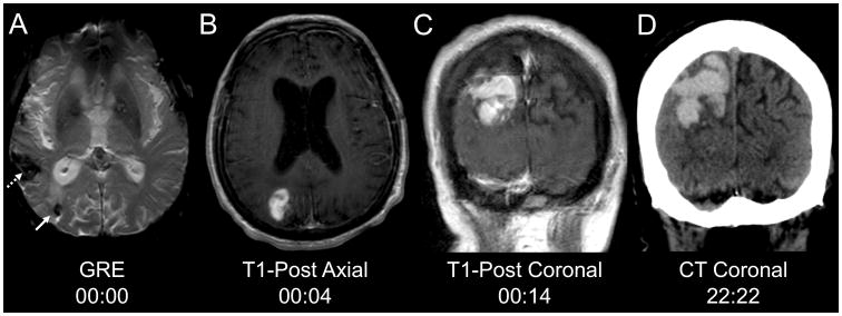
Fig. 1.
Hyperacute hemorrhage onset and expansion. a The chronic right temporoparietal hemorrhage (dotted arrow) and a new (since an MRI two years prior) right parietal microbleed (solid arrow) are seen on MRI gradient-recalled echo (GRE) sequence. b Four minutes later, contrast extravasation is present on the T1-post axial sequence. c Ten minutes later, the region of contrast extravasation has expanded on the T1-post coronal sequence. d A computed tomography scan 22 hours and 22 minutes after the GRE sequence demonstrates a large right parietal hyperdense lesion, consistent with hemorrhage.

