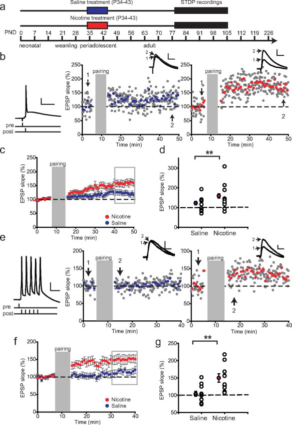
Figure 3.

Nicotine exposure during adolescence leads to a lasting increase in LTP in adult rats. a, Schematic representation of the experimental setup. Rats were treated with nicotine (red bar)/saline (blue bar) during adolescence, and the electrophysiological recordings took place starting 5 weeks after treatment. b, Example experiments showing STDP recorded from rats 5 weeks after treatment with saline (left panel) or nicotine (right panel) during adolescence using an induction protocol with single AP pairings. Induction protocol is shown left. The insets show example EPSP traces before (1) and after pairing (2); symbols and calibration are as in Figure 2. c, Average STDP in saline-treated rats (blue circles; n = 23 cells from 11 rats) and nicotine-treated rats (red circles; n = 22 from 10 rats). d, Summary of STDP results shown in c. The filled circles represent average EPSP slopes of last 10 min of the recordings (shown in gray rectangle); the open circles show individual experiments. **p < 0.01. Data are mean ± SEM. e, Example experiments showing STDP recorded from rats 5 weeks after treatment with saline (left panel) or nicotine (right panel) during adolescence using an induction protocol with a burst of five APs (left). f, Average STDP in saline-treated rats (blue circles; n = 11 cells from 5 rats) and nicotine-treated rats (red circles; n = 10 cells from 5 rats). g, Summary of STDP results shown in f. The filled circles represent average EPSP slopes of last 10 min of the recordings (gray rectangle); the open circles show individual experiments. **p < 0.01. Data are mean ± SEM.
