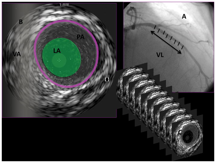
Figure 1. The methods for conducting 3-dimensional intravascular ultrasound (IVUS) examinations and definition of IVUS measurements.
IVUS was performed during routine coronary angiography with mechanical pullback (0.5 mm/s) from the mid to distal left anterior descending coronary artery to the left main coronary artery. The vessel length (VL), which was interrogated for each examination, was measured (A). The semiautomated contour software detected both the blood-media interface defined as lumen area (LA) and the media-adventitia interface defined as vessel area (VA). Plaque area (PA) was defined as the difference between VA and LA for each 2-dimensional image (B). Border detection was corrected manually in all frames after automatic border detection. Two-dimensional interrogations were performed at intervals of either 16 or 32 frames, depending on the heterogeneity of the image (C). Next, the vessel volume (VV), lumen volume (LV), and plaque volume (PV; mm3) were calculated with the Simpson rule for volumetric measurement and corrected for the segment length (mm3/mm). Plaque index (PI) was calculated as follows: PI=(PV/VV)×100%.

