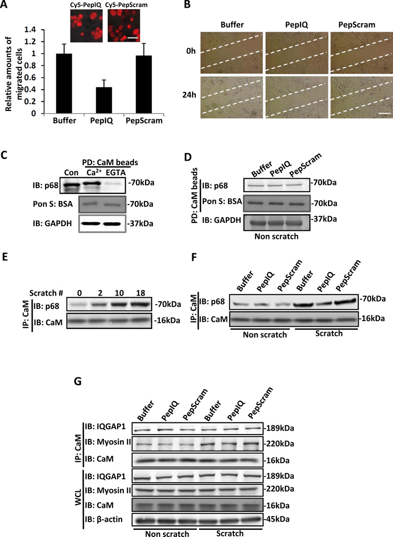
Figure 2. Cell migration and p68 - calmodulin interactions.
Cell migration of SW480 cells was analyzed by Boyden chamber assay (A) and monolayer scratch wound assay (B). Cells were treated by the indicated peptides (10 µg/ml). The upper two panels in (A) are representative confocal images of SW480 cells that were treated by the indicated Cy5.5-conjugated peptides, indicating peptide uptake. In (A), the number of cells migrating to the lower chamber is presented relative to the number of migrating cells that were treated by buffer. (C) Interaction of p68 with CaM in the presence of 2 mM CaCl2 (Ca2+) and 5 mM EGTA (EGTA) was probed by CaM pull-down (PD:CaM beads) followed by immunoblot using antibody against p68 (IB:p68). Cons are the control immunoblot of the cellular extracts using indicated antibody without CaM beads pull-down. (D) Interaction of p68 with CaM in extracts of SW480 cells was probed by CaM pull-down (PD:CaM beads) followed by immunoblot using antibody against p68 (IB:p68). The cells were treated by indicated peptides. Immunoblot of GAPDH (IB:GAPDH) is a loading control. (E) p68 and CaM interaction in extracts prepared from SW480 cells that were subjected to multiple scratch-wound treatment (number of scratches was indicated) analyzed by immunoprecipitation of CaM (IP:CaM). (F) p68 and CaM interaction in extracts prepared from SW480 cells that were subjected to 18 scratch wounds (Scratch) or no scratch wound (Non scratch), analyzed by immunoprecipitation of CaM (IP:CaM). Cells were treated by the indicated peptides. (G) CaM-IQGAP1 and CaM-Myosin II interactions in extracts prepared from SW480 cells that were subjected to 18 scratch wounds (Scratch) or no scratch wound (Non scratch), analyzed by immunoprecipitation of CaM (IP:CaM). The cells were treated by the indicated peptides. Immunoblots of IQGAP1 (IB:IQGAP1), Myosin (IB:Myosin II), and CaM (IB:CaM) in the whole cell lysate (WCL) indicate the cellular levels of these three proteins. Immunoblot of β-actin (IB:β-actin) in whole cell lysate is a loading control. The scale bars in A represent 20 µm and in B represent 100 µm.

