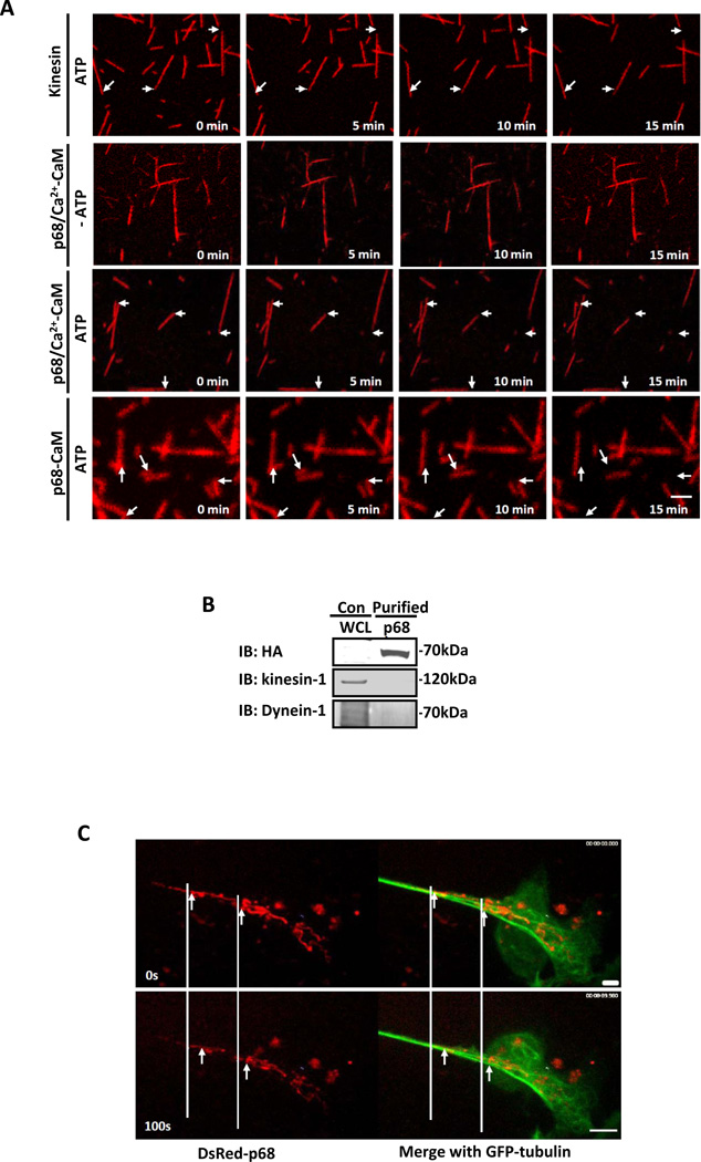
Figure 5. p68-CaM has microtubule motor activity.
(A) The movements of Rhodamine labeled microtubules on the glass slides on which the immunopurified HA-p68 (from SW480 cells) or bacterially expressed His-p68-CaM (p68-CaM) was attached in the presence (ATP) and absence (−ATP) and in the presence of 0.5 mM CaCl2 were recorded by the time lapse photography with confocal fluorescence microscopy (for video see on-line supplementary). The HA is a negative control with the HA peptide attached to the glass slides. (B) Existence of Kinesin-1 and Dynein-1 in the HA-p68 immunopurification was analyzed by immunoblots of the purified HA-p68 (IB:HA) using antibodies against kinesin-1 (IB:kinesin-1) and Dynein-1 (IB:Dynein-1). (C) Movements of DsRed-p68 along eGFP labeled microtubules in SW480 cells that were treated by scratch-wound were recorded by time lapse photography with confocal fluorescence microscopy (for video see on-line supplementary). In (A), arrows indicate the same positions of different time points. In (C), the arrows indicate the moved DsRed-p68. The scale bars in A and C represent 5 µm.

