Fig. 6.

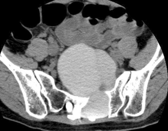
CT myelography in case 10 showing the huge cyst (supine position, caudal view). The left S2 root is compressed and the S3 root is continuous with the cyst interior

CT myelography in case 10 showing the huge cyst (supine position, caudal view). The left S2 root is compressed and the S3 root is continuous with the cyst interior